Epilepsy & Seizures
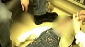
Tonic status epilepticus
Jan. 20, 2025
MedLink®, LLC
3525 Del Mar Heights Rd, Ste 304
San Diego, CA 92130-2122
Toll Free (U.S. + Canada): 800-452-2400
US Number: +1-619-640-4660
Support: service@medlink.com
Editor: editor@medlink.com
ISSN: 2831-9125
Toll Free (U.S. + Canada): 800-452-2400
US Number: +1-619-640-4660
Support: service@medlink.com
Editor: editor@medlink.com
ISSN: 2831-9125
Worddefinition
At vero eos et accusamus et iusto odio dignissimos ducimus qui blanditiis praesentium voluptatum deleniti atque corrupti quos dolores et quas.
Reflex anoxic seizures are a common type of nonepileptic seizure mainly encountered in infants and younger children but occasionally persisting into adulthood. They are dramatic and frightening to witness but in nearly all cases benign and without sequelae. They are often confused with other forms of syncope. Exceptionally, they may trigger epileptic seizures. This phenomenon has been reported in adults as well as in children. In this article, the author reviews their clinical manifestations, discusses their differential diagnosis (concentrating on the need to consider rare but potentially fatal conditions that may mimic reflex anoxic seizures, such as long QT syndromes), and considers the latest approaches to management.
• Reflex anoxic seizures are a form of syncope usually starting in infancy or early childhood. | |
• They are precipitated by noxious stimuli, which can be physical or emotional. | |
• For reasons yet unknown, the noxious stimulus causes a vagally mediated brief asystole, which is responsible for the syncope. | |
• The clinical manifestations are dominated by the child losing awareness and falling to the ground, appearing a deathly white color. Convulsive features are common. | |
• Reflex anoxic seizures are benign in the sense that sufferers always recover from them. If they are very frequent, cardiac pacing can be helpful. | |
• Reflex anoxic epileptic seizures may occur with pathogenic variants, such as SCN1B or SCN8, but the neurodevelopment is abnormal in these cases. |
Reflex anoxic seizures are a form of syncope encountered mainly, but not exclusively, in young children. A seizure is, in the broadest sense of the word, any clinical event caused by a transient (but not necessarily short-lived) alteration of cerebral function. When the seizure is epileptic, it is caused by an epileptic mechanism, ie, as a consequence of excessive or hypersynchronous discharge of cortical neurons. By definition, “syncope” is from the Greek, “a cutting off,” implying an abrupt interruption in the supply of energy to the cerebral cortex. This is usually a consequence of a reduction in cerebral perfusion by oxygenated blood. It can be a result of either a sudden reduction in the blood flow to the brain, a drop in the oxygen content of the blood supplying the brain, or a combination of the two. It follows that syncope can be a consequence of numerous different pathophysiological mechanisms but that they are nonepileptic because they do not arise as a consequence of excessive or hypersynchronous discharge of cortical neurons. Moreover, syncope can have different clinical manifestations, ranging from transient loss of consciousness, usually accompanied by a decrease or loss in postural tone (the principal manifestations of “simple faints”), to tonic and myoclonic events and nonepileptic spasms.
To avoid confusion, it is perhaps best to consider the terms “anoxic seizure” and “syncope” as being synonymous and meaning clinical events caused by transient alterations in cerebral function arising as a consequence of sudden reductions in cerebral perfusion of oxygenated blood. Numerous types of anoxic seizure or syncope have been described. The best known, if not necessarily the best understood, is the “simple faint” or vasovagal syncope. At least in infants and children, breath-holding attacks and reflex anoxic seizures are also widely recognized and common causes of anoxic seizures or syncope. Other types include vagovagal syncope, cardiac syncope (including long QT disorders, other cardiac arrhythmias, and structural cardiac disease), syncope due to orthostasis, hyperventilation, compulsive Valsalva maneuvers, gastroesophageal reflux, and imposed upper airway obstruction (suffocation). In addition, anoxic seizures or syncope are a feature of both hyperekplexia and familial rectal pain syndrome. Finally, there are likely to be other types of anoxic seizure or syncope not yet characterized.
Reflex anoxic seizures are a particular type of anoxic seizure or syncope, most commonly seen in young children in whom an anoxic seizure or syncope is provoked or precipitated by a noxious stimulus (hence, “reflex”). Various precipitants have been identified, but the most common is an unexpected bump to the head. Breath-holding attacks have been recognized for centuries. However, it is only relatively recently that their pathophysiology has begun to be understood, and in consequence, their separation from reflex anoxic seizures has been recognized. Indeed, the distinction between the two may not be complete.
Stephenson reviewed the historical development of the concept of breath-holding attacks and reflex anoxic seizures and the separation of the two (37). Key developments were:
(1) Clear description by Bridge and colleagues of the typical features of mild breath-holding spells (03). | |
(2) Description of the clinical and EEG features of anoxic convulsions in children by Gastaut and Gastaut (09). | |
(3) Anoxic anoxia proposed as the mechanism of breath-holding spells by Gauk and colleagues (10). | |
(4) Description by Maulsby and Kellaway of the clinical features during more severe attacks, including the occurrence of a convulsive phase (20). | |
(5) Separation by Maulsby and Kellaway of anoxic seizures into two types: Type I, corresponding to (blue) breath-holding spells, and type II (hypoxic crises) (20). | |
(6) Confirmation by Lombroso and Lerman that two distinct types of breath-holding spells could be distinguished (18). These authors preferred the terms cyanotic breath-holding and pallid infantile syncope. However, they also demonstrated that both types of attack could occur in the same child. | |
(7) The term “reflex anoxic seizure” proposed by Stephenson for the type II spells of Maulsby and Kellaway and the pallid infantile syncope of Lombroso and Lerman (33). This term has now been widely adopted. | |
(8) Recognition that true epileptic seizures can exceptionally follow reflex anoxic seizures in children (anoxic-epileptic seizures) and in adults (36; 37; 13). | |
(9) Reflex anoxic seizures may occur with de novo pathogenic SCN8A variants, but the neurodevelopment is abnormal in these cases (26). |
It is now generally recognized that breath-holding attacks and reflex anoxic seizures represent distinct pathophysiological phenomena, which, nevertheless, can occur in the same subject. It has become clear that prolonged respiratory apnea is a principle, if not the only, mechanism responsible for breath-holding spells in which cyanosis is a major feature and that brief cardiac asystole as a consequence of excess activity of the vagus nerve is the cause of reflex anoxic seizures. This may also be the case with de novo pathogenic SCN8A variants (26).
Reflex anoxic seizures occur in otherwise normal children, although there is no reason to suppose that children with disorders such as cerebral palsy and mental retardation are protected from them. They usually start in infancy or early childhood. Presumably because the precipitants to the attacks generally require a degree of mobility, descriptions of reflex anoxic seizures before the age of 6 months are rare. However, Stephenson describes a case with onset at 4 weeks of age (case 9.39) (37). I have personally seen a case in which attacks of anoxic seizures accompanied by cardiac asystole (ie, identical to reflex anoxic seizures) occurred in the context of familial rectal pain syndrome and started on day 1 of life. Lombroso and Lerman state that in 15 out of 193 subjects, attacks started in the neonatal period (18); however, they did not distinguish whether these were breath-holding attacks or reflex anoxic seizures. DiMario reported that 5% of breath-holding spells started in the perinatal period, and as early as within hours of birth in a single case but did not distinguish reflex anoxic seizures from other types of breath-holding spells (06). Only 3% of subjects started to have attacks between 25 and 30 months of age. However, there are many descriptions of attacks starting in later childhood and in adult life, although in such cases, the precipitants tend to be different, for example, involving bloodletting (27) or dental extractions.
A minor bump to the head is the most commonly reported precipitant. Usually the toddler trips and falls; the child’s caregiver may hear the bump. Most commonly, the child does not cry, although some parents give descriptions of the child “trying to cry” (33), or there may be a gasp or a sob. Syncope rapidly ensues. Indeed, the short latency between the stimulus and the attack has been emphasized as an important distinction from the more familiar (at least in older children and adults) vasovagal syncope. The child loses awareness and postural tone, falling to the ground. There may be downbeat nystagmus. The child is likely to be pale, sometimes described as “deathly white,” which is entirely appropriate given that they are likely to be asystolic; however, it is important to note that not all children go pale (or at least are perceived as going pale by their caregivers).
Stephenson records descriptions from parents of “blue or purple lips,” “yellow patches through the blue,” and of no noticeable color change (33). In some attacks, the child rapidly returns to normal following the limp or pallid phase. However, more usually there is a convulsive phase. This is usually manifested with tonic stiffening, often amounting to opisthotonus, and often includes clenching of the jaw and hands. Video recordings of other forms of anoxic seizures (vasovagal syncopes) suggest that there may be marked asymmetry (17). Parents may report the eyes to have rolled or to be “popping out of the head.” A few clonic jerks of the limbs or spasms are often noted. Urinary incontinence is not uncommon (33). Any initial limpness may be so short that the whole attack is dominated by the convulsive components.
Recovery is often rapid, but usually the child is sleepy after the attack, and there may be persisting pallor. Lombroso and Lerman reported that the length of the postictal stupor reflected the duration of the asystole up to a maximum of 3 minutes of stupor (18). Some cases reported by Stephenson took longer to recover (37).
Although minor bumps to the head are reported as the most common precipitants to reflex anoxic seizures, many other stimuli may also be involved. Lombroso and Lerman emphasized the importance of minor injuries and sudden fright (18). They noted that occipital blows to the head appeared to be particularly provocative. Stephenson reported that pain, especially from emotion (surprise, fear, annoyance, frustration, and excitement), crying, and fever were provocative factors (33). Fever was reported as a provocative factor in 14% of cases. Some cases of fever-induced reflex anoxic seizures are likely to be misdiagnosed as febrile (epileptic) seizures, as has been emphasized by a number of authors. Many, if not most, cases of venipuncture fits are reflex anoxic seizures. When one considers the vast range of situations in which a child (or adult) can be surprised, frightened, upset, or merely excited, it is easy to understand how reflex anoxic seizures can occur in special settings, such as bathing and water immersion; in the anesthetic room; when witnessing “blood and gore”; at the dentist office, school, place of worship, or the hairdresser’s; and whilst watching television as described by Stephenson (37).
The precipitants and the manifestations of reflex anoxic seizures may change with age. Hence, in unsteady toddlers, minor bumps to the head are likely to predominate, whereas in the older child, adolescent and adult factors such as the sight of blood or venipuncture are likely to be more relevant. The adult physician is likely to classify such events as vasovagal syncopes rather than as reflex anoxic seizures and indeed progression through reflex anoxic seizures to vasovagal syncope is recognized. In this regard, Stephenson and McLeod note that beyond the toddler stage, children with reflex anoxic seizures may report out-of-body experiences with a dream-like quality (39). In older age groups, reflex anoxic seizures can occur with acupuncture or dry needling (43).
There is considerable variation in the frequency of reflex anoxic seizures. Some subjects undoubtedly only ever have a single attack whilst other well-documented cases have multiple daily attacks. The attacks have been reported to generally reach a peak in frequency towards the end of the first or beginning of the second year of life.
Distinguishing epileptic from nonepileptic seizures has, historically, been an important stage in the evolution of our understanding of paroxysmal events. It is, therefore, somewhat unsettling that, following decades when the importance of the separation of epileptic from nonepileptic seizures was stressed, strong evidence has emerged that exceptionally a reflex anoxic seizure (and also other syncopes such as cyanotic breath-holding attacks and compulsive Valsalva maneuvers) can trigger brief myoclonic jerks and an epileptic seizure (anoxic-epileptic seizure). In the modern era, it was Stephenson who first brought this to attention (36; 37). However, there appear to be examples in the cases reported by Bridge and colleagues (03), and other authors have also reported examples (01). Published cases have included EEG recordings in which cardiac asystole is followed first by the characteristic slowing and flattening of the EEG and then by ictal epileptiform discharges, the latter being accompanied by clinical manifestations such as atypical absences or myoclonic or clonic seizures. The myoclonic jerks may also occur with the reperfusion of the brain without frank epileptiform discharges (Moshé, personal observations). Seizures, considered by the authors to be generalized tonic-clonic and status epilepticus, following breath-holding attacks have also been reported (15). Stephenson and coworkers have reported cases with home video recordings (38). True epileptic seizures triggered by syncopes have been described in adults (13).
All authors stress that, in the absence of an identified cause (such as a pathogenic SCNA8 variant) or significant comorbidity (cardiovascular disease), reflex anoxic seizures are benign. They are not associated with an increased mortality, and they do not cause cardiac or cerebral damage. A reported case of a fatal cardiac arrest as a consequence of pallid syncope is not wholly convincing (40).
Lombroso and Lerman reported that the attacks are spontaneously outgrown by school age in the vast majority of instances (18). DiMario reported that in those children with pallid breath-holding spells who had achieved a 1-year remission, the median age at remission was between 37 and 42 months (06). STARS, a UK based self-help group for children and families with reflex anoxic seizures, state on their web site that unpublished data (from a patient and family survey) suggest that 75% of children outgrow the attacks by school age (5 years of age in the UK) but that they continue into adult life in some cases (STARS web site).
It is accepted that children with reflex anoxic seizures have a higher incidence of subsequent vasovagal syncope.
Reflex anoxic seizures are not epileptic in origin.
The fundamental cause of reflex anoxic seizures is not known.
Although it is established that reflex anoxic seizures arise as a consequence of a vagally mediated asystole, the cause of this is unknown. It is usually explained on the basis of a phasic increase in vagal tone. Presumably, either increased firing of the vagal nerves in response to a particular stimulus or increased sensitivity of the heart to the effects of the vagal nerves could give rise to reflex anoxic seizures. Increasingly, channel ion defects are being shown to be important causes of cerebral and extracerebral paroxysmal episodes. However, to date, such a mechanism has not been demonstrated in reflex anoxic seizures.
The principal event in the production of a reflex anoxic seizure is asystole. The corresponding reduction in the delivery of blood and oxygen to the brain is the basis of the syncope. The motor features, so common in reflex anoxic seizures, are probably some form of release phenomena mediated at the level of the brainstem, temporarily freed from higher cortical control as a consequence of the ischemia.
The asystole responsible for the cerebral ischemia is attributed to increased vagal input to the heart (vagal input exerting a cardioinhibitory effect) triggered by the various stimuli identified as important in the production of reflex anoxic seizures.
The ocular compression testing has been important in increasing our understanding of reflex anoxic seizures (35). The test relies on the oculocardiac reflex, which is a brainstem reflex mediated via the ophthalmic division of the trigeminal nerve and the vagus. Ocular compression in normal subjects characteristically gives rise to a vagally mediated increased R-R interval on the ECG. Subjects with reflex anoxic seizures characteristically show a much-reduced latency in the first measurable R-R interval increase and 6 seconds or more of asystole. Moreover, the test often precipitates habitual attacks. However, this testing is rarely carried out nowadays. Tilt-testing may also be abnormal in a minority of children with reflex anoxic seizures beyond about 7 years of age. Genetic testing can be carried out if there are significant additional neurodevelopmental abnormalities.
There have been no recent epidemiological studies on the subject. Lombroso and Lerman reported in the 1960s a history of breath-holding spells in 4.6% of children, of whom 62% had cyanotic infantile syncope, 19% had pallid infantile syncope (corresponding to reflex anoxic seizures), and 19% had features of both (18). This would imply a prevalence in the population studied (children from birth to 8 years of age whose mothers were normal, registered in the Maternal and Infant Health Collaborative Project, and delivered at the Boston Lying-In Hospital) of 0.8% to 1.7%
Lombroso and Lerman reported a family history of breath-holding attacks to be twice as common as in controls (23% vs. 11%) (18). Unfortunately, it is not possible to compute separate figures for the different types of breath-holding. DiMario identified a positive family history in 34% of cases of cyanotic and pallid breath-holding spells (06).
Hindly and colleagues found that the diagnosis was syncope in 42% of 380 children referred to a secondary care “fits, faints, and funny turns” clinic in the UK. In 54 children, the mechanism of syncope was considered to have been vagal (12).
Reflex anoxic seizures can be prevented or their frequency reduced if the precipitants to the attacks are avoided. This may be impractical in many cases.
Reflex anoxic seizures need to be distinguished from epileptic seizures (including febrile seizures) and from other syncopes.
Epileptic seizures. The same triggers that characteristically trigger reflex anoxic seizures can exceptionally trigger epileptic seizures. However, so rare are such cases that the principle means of distinguishing reflex anoxic seizures from epileptic seizures is the circumstances of the attacks. This should be easy when a clear history is available. Unfortunately, in many cases, particularly if only one or a few attacks have occurred, the events leading up to the attacks may not have been witnessed or accurately recalled. The clinical manifestations of reflex anoxic seizures may suggest epileptic drop attacks. However, drop attacks rarely occur in otherwise normal infants and children, whilst reflex anoxic seizures do. Also, pallor is not a common feature of epileptic drop attacks. When reflex anoxic seizures are accompanied by particularly prominent convulsive features, they may be misinterpreted as generalized tonic-clonic seizures (either primarily or secondarily generalized). However, the characteristic sequence seen in reflex anoxic seizures of collapse with pallor, followed later by stiffening or jerking, would be unusual even for a secondarily generalized tonic-clonic seizure. As previously noted, true epileptic seizures can rarely be triggered by reflex anoxic seizures.
Breath-holding attacks. There has been much confusion regarding the relationship between breath-holding attacks and reflex anoxic seizures. The terms are sometimes used interchangeably. It is now common (but not universal) to distinguish breath-holding attacks (sometimes called blue or cyanotic breath-holding attacks) from reflex anoxic seizures (sometimes called white or pallid breath-holding attacks), whilst recognizing that some children appear to have both and that some attacks appear to have features of both (sometimes called mixed breath-holding attacks). Although the term “breath-holding” suggests a voluntary behavioral etiology, this is not the case. Breath-holding attacks usually follow a painful or emotional stimulus that causes the child to cry. After a brief period of time, the breath appears to be held in expiration, with loss of consciousness and color change (often deep cyanosis, hence, the name). The child is initially floppy, but as in reflex anoxic seizures, opisthotonus and other convulsive phenomena may ensue. The pathogenesis of breath-holding attacks is still incompletely understood. A major component appears to be prolonged expiratory apnea, without appreciable change in cardiac rate or rhythm (30). Unlike the situation in many syncopes, particularly those familiar to adult physicians, hypotension is not a feature. Indeed, arterial hypertension has been demonstrated (10). Syncope in these attacks should be considered to arise from hypoxic hypoxia (anoxic anoxia), rather than to reduced cerebral perfusion. It has been suggested that the term “prolonged expiratory apnea” should replace breath-holding attacks. However, as previously noted, cases are described in which there is both prolonged expiratory apnea and bradycardia or asystole. The role of a forced Valsalva maneuver in the pathogenesis of breath-holding attacks has also been suggested but is less well supported evidentially. Moreover, Gauk and colleagues reported a case occurring in a subject without a glottis (aglottic breath-holding spells) (11).
Vasovagal syncope (simple faints). This is the commonest cause of syncope overall and occurs in all ages. There is overlap in the triggers, the clinical features, and the underlying pathogenesis of both vasovagal syncopes and reflex anoxic seizures—and also (cyanotic) breath-holding attacks—both being examples of neurally mediated (or neurocardiogenic) syncope. The major difference pathogenetically is that in reflex anoxic seizures, the main effect is a vagally induced asystole, whereas in vasovagal syncope, a major vasodepressor effect with decreased sympathetic tone leading to vasodilation and, hence, hypotension occurs with a variable vagally mediated reduction in heart rate (05). Stimuli causing vasovagal syncopes can usually be identified but may be subtle. Pain, fear, and other noxious stimuli are important triggers. In children they often occur after baths and when getting their hair blow dried. Although they commonly occur in the upright position, it is a common fallacy to consider them excluded if one or more attacks occur while sitting or supine. In vasovagal syncope, at least in older children and adults, prodromal symptoms (often called pre-syncope) are usually, but not always, described. These include nausea, lightheadedness, blurred vision, auditory hallucinations (usually ringing noises), feelings of warmth, and abdominal pain. Witnesses often describe pallor. As in reflex anoxic seizures, the subject loses consciousness and is initially floppy. However, convulsive features are again common. Some children show a transition from reflex anoxic seizures in infancy to vasovagal syncope later on.
Vagovagal syncope. These are rare. Both the afferent and efferent limbs are vagally mediated, the former often being triggered by swallowing or vomiting, leading to a vagally mediated cardiac inhibition.
Gastroesophageal reflux. Reflux of gastric contents up the esophagus is common in normal babies and infants and especially so in those with neurologic impairments such as cerebral palsy. A number of clinical pictures are described. Sandifer syndrome is characterized by contortions of the neck and is not likely to be confused with reflex anoxic seizures. The awake apnea syndrome consists of attacks occurring within an hour of a feed, often following a change in posture (31). A gasp is followed by apnea, color change, and often stiffening. Such attacks may be a form of vagovagal syncope, but laryngeal spasm may also be involved.
Cardiac syncopes. The principle clinical manifestations of reflex anoxic seizures are all a consequence of cardiac standstill, notwithstanding the fact that they are neurally mediated rather than reflecting a primary cardiac disorder. It is, therefore, not surprising that they may be indistinguishable from syncopes due to a variety of cardiac disorders, whether structural or caused by cardiac dysrhythmias. In children with known heart disease, diagnostic difficulties are unlikely. However, one of the most important groups of conditions to be considered in any child presenting with attacks consistent with reflex anoxic seizures are the long QT disorders. These include Lange-Nielsen syndrome (autosomal recessive with congenital deafness) and Romano-Ward syndrome (autosomal dominant with incomplete penetrance). Children and adults with long QT syndromes are prone to episodes of life-threatening ventricular tachyarrhythmias, especially torsades de pointes. The same triggers that give rise to reflex anoxic seizures have been reported in one case (02) to have triggered a ventricular tachyarrhythmia in association with a prolonged QT. However, attacks are particularly likely to occur during exercise and also during sleep. In most instances, these disorders can be diagnosed by demonstrating a prolonged QT interval on a standard EEG. However, sometimes exercise testing may be required, and when there is any suspicion of such a disorder, cardiological assessment should be obtained. Molecular DNA testing is likely to assume an increasingly important role because prolonged QT is associated with mutations in genes coding for sodium and potassium ion channels.
Imposed upper airway obstruction. Those in charge of babies and infants (predominantly mothers) may, sometimes as a manifestation of Meadow syndrome, suffocate them, for example by a hand, pillow, or bosom (23). For obvious reasons, the initial stages of such episodes are rarely witnessed, except if there is covert videorecording. The subsequent description offered by the perpetrator or other witnesses called to see the later stages of the attack may strongly suggest a reflex anoxic seizure. A high degree of suspicion is required if all the attacks occur in the presence of a single individual, especially if no one else has witnessed the beginning of the attack but the collapsed infant has been shown to other relatives or professionals before recovery (28).
Syncopes in association with posterior fossa abnormalities. Children with posterior fossa anomalies, such as Chiari malformations, may develop syncopes with features similar to reflex anoxic seizures. Attacks may be precipitated by stimuli that raise intracranial pressure, such as coughing.
Syncopes in children with learning difficulties or mental retardation. Some such children repeatedly induce syncopal attacks, which may have a superficial resemblance to reflex anoxic seizures, by maneuvers such as hyperventilation followed by forced Valsalvas.
Hyperekplexia. This dominant or occasionally recessive disorder caused by glycine receptor mutations is often characterized by attacks in which the predominant feature is tonic stiffening. These may be provoked by anything causing a startle and may be life-threatening.
Paroxysmal extreme pain disorder (previously familial rectal pain syndrome). In this rare, dominant disorder of autonomic control, excruciating attacks of pain occur spontaneously or in response to various stimuli, including defecation and eating. It is due to mutations on the SCN9A sodium ion channel. Attacks indistinguishable from reflex anoxic seizures may occur. However, their onset is within the first few days of life, the triggers usually include defecation, and there is associated flushing and harlequin color changes (08).
Reflex anoxic seizures occur in otherwise healthy subjects.
The diagnosis of reflex anoxic seizures is primarily a clinical one, made on the history. If this is typical and there are no abnormal physical findings, then no further investigations are required. However, if there are any atypical features, particularly a family history of early sudden, unexplained death or a history in the patient of attacks during exercise or when asleep, a prolonged QT disorder must be excluded. In the first instance, a standard ECG should be obtained and the QT interval measured and compared against normal values. Even if this is normal, if there remains doubt, referral for a cardiological opinion is justified because the QT interval is not always prolonged on a routine ECG. Many clinicians now consider it justified to measure the QT interval in all children having seizures or syncopes because of the importance of not missing a prolonged QT disorder.
There is evidence that in some children with breath-holding attacks correction of concomitant iron deficiency anemia may reduce the frequency of attacks. This appears to relate to blue breath-holding attacks rather than reflex anoxic seizures. However, as there is an overlap, it may be justified to perform a full blood count and to measure serum ferritin.
If the diagnosis is in doubt, it is likely to be useful if one or more attacks can be viewed. If they are frequent, the parents may be able to record one or more on a camcorder.
Routine EEG is generally not helpful. Indeed if a diagnosis of probable reflex anoxic seizure is made, obtaining an EEG may be misleading. Reflex anoxic seizures are not associated with interictal EEG abnormalities. Given that otherwise normal children may have abnormal interictal EEGs, an EEG might show features that are irrelevant to the child’s attacks but that will be misinterpreted as evidence of an epileptic origin of the attacks.
Occasionally it may be justified to obtain an ictal EEG recording in a child who is experiencing attacks, the nature of which is not clear. In babies and infants who are having apneic attacks of obscure origin, polysomnography may be helpful. This should ideally include EEG, ECG, and EMG leads, a respiratory strain gauge (to detect respiratory movements), a thermistor placed at the nostrils (to detect airflow), an oxygen saturation monitor, and an esophageal pH probe. Simultaneous video recording is likely to be helpful. Using such a system, it should be possible to determine the nature of the apneas in most cases. In an older child who is having frequent attacks and whose etiology is unclear, an ambulatory “cassette” EEG may be an appropriate investigation. However, additional useful information is likely to be obtained if any attacks can also be viewed, arguing in favor of video-telemetry. Finally, an ictal EEG recording may be obtained as part of an ocular compression test (discussed below).
However obtained, the EEG during a reflex anoxic seizure will show characteristic abnormalities that are due to hypoxia.
These were first described by Gastaut and Gastaut and consist of an orderly series of changes (09):
(1) Desynchronization (not always obvious) |
The EEG changes first appear 3 to 8 seconds after the beginning of asystole. It should be noted that these changes are not specific for reflex anoxic seizures; they are seen in all syncopes associated with hypoxia. Minor differences that may occur in different types of syncope are reviewed by Stephenson (37).
The ocular compression test has been advocated by some as a useful confirmatory test for reflex anoxic seizures, whereas others consider it unnecessary, even dangerous. It depends on the oculocardiac reflex, a brainstem reflex involving the ophthalmic division of the trigeminal nerve as the afferent limb and the vagus as the efferent. Activation of the reflex results in an increase in the R-R interval. In children with reflex anoxic seizures, the reflex is exaggerated, often inducing asystole and reflex anoxic seizures. Different methodologies have been used. Stephenson suggests pressing the eyes (under the closed eyelids, below the supraorbital ridges) firmly backwards, using enough pressure to blanch the thumbnail beds for 2 millimeters and maintaining this for exactly 10 seconds. During the test, both an EEG and ECG should be running. Normative data, summarized by Stephenson, indicate that few (0% to 4%) normal children will have asystole lasting longer than 6 seconds (37) during ocular compression. The asystole is said to nearly always begin during the 10 seconds of ocular compression, but occasionally a tardive response is seen, with the asystole beginning a few seconds after the end of the ocular compression.
Concerns have been raised about the risks involved, including ocular trauma (especially in high myopia and glaucoma) and psychological trauma. Stephenson has defended the test as without complication when used in the awake child in the investigation of suspected reflex anoxic seizures. Most children with reflex anoxic seizures can be diagnosed without undertaking the ocular compression test. However, it may be useful in the investigation of subjects with atypical attacks.
Head-up tilt testing has also been used in the evaluation of children with reflex anoxic seizures. However, it is generally considered unsuitable for children under the age of 10 years. It is now extensively used in the evaluation of syncopes, especially when vasovagal attacks are suspected. In normal subjects, tilting causes a reduction in venous return that is compensated by increased adrenergic tone. In subjects with vasovagal syncope, tilting causes decreased venous return, leading to increased sympathetic tone with stimulation of cardiac C fibers. This causes a vasodepressor effect mediated at brainstem level, resulting in a sudden reduction in sympathetic tone (vasodilation) and increase in vagal tone (bradycardia) and, hence, syncope (05). Tilt-testing is not generally considered suitable for younger children, which obviously limits its usefulness in a condition usually starting in infancy. However, when used in older children with reflex anoxic seizures, asystole is frequently provoked (37).
The ECG changes during reflex anoxic seizures are characteristic. Therefore, prolonged ECG recordings can also be useful in cases of diagnostic difficulty. Depending on the frequency of attacks, ambulatory 24-hour EEG (Holter monitoring), patient-activated event recorders, and even (if the situation justifies it) implantable loop recorders can be useful. A good example of the use of a cardiac event recorder is provided by McLeod and colleagues (21). Indeed, there is a major effort to develop wearable technologies to improve patient care and overall management (16).
The most important aspect of management is detailed explanation to the parents about the condition and reassurance that it is a benign disorder that is often, but not always self-limiting. Written and web-based information is usually greatly appreciated. With regards to the latter, the website of STARS (www.stars.org.uk) can be strongly recommended. Caregivers need to be told that they will not be required to resuscitate their child. It is often recommended that children should be placed in the recovery position during or after reflex anoxic seizures. Whether this is of value remains to be determined.
Given the pathophysiology of reflex anoxic seizures, they would be expected to respond to treatment with anticholinergic drugs such as atropine. The literature is limited regarding the use of such drugs to treat reflex anoxic seizures. Success has been reported with oral atropine methonitrate, oral atropine sulphate, and transdermal scopolamine (34; 22; 25). With regards to the atropine preparations, the methonitrate is preferred to the sulphate because of its reported extracerebral distribution, eliminating concerns about adverse effects on cognitive function. However, it may be difficult to obtain. Oral preparations of atropine generally need to be given frequently (three to five times a day). The reported daily dose necessary to obtain control varied in a series reported by McWilliam and Stephenson from 50 to 240 µg/kg/dose (22). Side effects are predictable and include dry mouth and blurred vision. Overdose can be fatal. Because of the benign nature of reflex anoxic seizures and the problems of pharmacological treatment, it is not generally used but is reserved for the most difficult cases.
Glycopyrrolate is an anticholinergic drug with a longer half-life than atropine. Carano and colleagues described its use combined with theophylline (because of its chronotropic and respiratory stimulating effect) to successfully treat a 13-month-old child with severe pallid breath-holding spells (04), and a further case report of its use has been published (41).
Treatment with oral iron preparations has been advocated for breath-holding spells. Mocan and colleagues reported that 63 out of 91 such children had evidence of iron deficiency anemia and were treated for this (24). After 3 months of treatment, there was a significant reduction in the number of spells in those who had been treated compared to those who had not. However, relatively few children had reflex anoxic seizures (pure pallid breath-holding spells), and it appears that only the cyanotic spells showed a clear response. A Cochrane review concluded that iron supplements significantly reduced the frequency of breath-holding attacks (OR 76.48; 95% CI 15.65 to 373.72; P< 0.00001) and that iron supplementation (5 mg/kg/day of elemental iron for 16 weeks) appeared to be useful in reducing the frequency and severity of breath-holding attacks, particularly, but not only, in those with iron deficiency anemia (44).
Permanent cardiac pacing as a treatment for reflex anoxic seizures was first reported in 1996 (32). Subsequently, a blinded study of its use in 12 children aged 2 to 4 years with frequent reflex anoxic seizures found a significant reduction in episodes of loss of consciousness during pacing (21). Six patients had no loss of consciousness when the pacemaker was programmed to sensing only; three patients had no further episodes of syncope whether paced or not; and two patients continued to have episodes of syncope despite being paced. Two patients reported unpleasant symptoms of dizziness and awareness of the pacemaker pacing. Infection prompted one pacemaker to be removed. It was concluded that permanent cardiac pacing is an effective treatment for frequent and severe reflex anoxic seizures that have not responded to medical treatment. Subsequently, there have been other reports of the successful treatment of reflex anoxic seizures with cardiac pacing (42; 07; 14; 29). However, in another study, the administration of intranasal midazolam (5 mg) one minute prior to venipuncture prevented the anticipated syncope that was induced previously under ECG and EEG monitoring (19). The authors suggested that anxiety may play a role.
Previously it was noted that in a small number of children, reflex anoxic seizure can trigger epileptic seizures (anoxic-epileptic seizures). The efficacy and appropriateness of antiseizure drug treatment in such cases has not been evaluated, but anecdotal evidence of efficacy has been published by Stephenson and colleagues (38). Ranza and colleagues reported two cases with a de novo pathogenic SCNA8 variant (26). One responded to carbamazepine, perhaps suggesting gain of function.
As explained above, the most important aspect of management is good communication with the parents or other caregivers.
Special thanks to Drs. Sameer Zuberi and Michael Blackburn for their help with illustrations.
All contributors' financial relationships have been reviewed and mitigated to ensure that this and every other article is free from commercial bias.

Solomon L Moshé MD
Dr. Moshé of Albert Einstein College of Medicine has no relevant financial relationships to disclose.
See ProfileNearly 3,000 illustrations, including video clips of neurologic disorders.
Every article is reviewed by our esteemed Editorial Board for accuracy and currency.
Full spectrum of neurology in 1,200 comprehensive articles.
Listen to MedLink on the go with Audio versions of each article.
MedLink®, LLC
3525 Del Mar Heights Rd, Ste 304
San Diego, CA 92130-2122
Toll Free (U.S. + Canada): 800-452-2400
US Number: +1-619-640-4660
Support: service@medlink.com
Editor: editor@medlink.com
ISSN: 2831-9125
Epilepsy & Seizures
Jan. 20, 2025
Epilepsy & Seizures
Jan. 09, 2025
Epilepsy & Seizures
Jan. 09, 2025
Epilepsy & Seizures
Dec. 23, 2024
Epilepsy & Seizures
Dec. 19, 2024
Neurogenetic Disorders
Dec. 15, 2024
General Neurology
Dec. 14, 2024
General Child Neurology
Dec. 10, 2024