- Clinical Categories
Infectious Disorders
-

Media
Three-dimensional structural view of the icosahedral-shaped poliovirus type 3 based on x-ray diffraction
(Source: Filman DJ and Hogle JM (1989) [https://www.rcsb.org/structure...]. Creative Commons Attribution-Share Alike 4.0 International [CC BY 4.0] license, creativecommons.org/licenses/by-sa/4.0/deed.en.)
-

Media
Internal auditory canal of a patient with herpes zoster oticus (MRI) (1)
Internal auditory canal MRI image of a patient with herpes zoster oticus. The post-contrast 3D-FLAIR MRI shows high-signal intensities of cochlea (long arrow), posterior semicircular canal, lateral semicircular canal (large arrow), and vestibule (arrowhead) in the right ear. (Source: Lee J, Choi JW, Kim CH. Features of audio-vestibular deficit and 3D-FLAIR temporal bone MRI in patients with herpes zoster oticus. Viruses 2022;14[11]:2568. Creative Commons Attribution 4.0 International [CC-BY 4.0] license, creativecommons.org/licenses/by/4.0.)
-

Media
Nonspecific findings on facial nerve conduction study in Ramsay-Hunt syndrome
A facial nerve conduction study revealed nonspecific findings. NCV = nerve conduction velocity. (Source: Hwang YS, Kim YS, Shin BS, Kang HG. Two cases of Ramsay-Hunt syndrome following varicella zoster viral meningitis in young immunocompetent men: case reports. BMC Neurol 2023;23[1]:43. Creative Commons Attribution 4.0 International [CC-BY 4.0] license, creativecommons.org/licenses/by/4.0.)
-

Media
Pure-tone audiometry revealing high-frequency sensorineural hearing loss in Ramsay-Hunt syndrome (1)
Pure-tone audiometry revealing both increased air and bone conduction thresholds without an air-bone gap in the right ear, consistent with high-frequency sensorineural hearing loss. (Source: Hwang YS, Kim YS, Shin BS, Kang HG. Two cases of Ramsay-Hunt syndrome following varicella zoster viral meningitis in young immunocompetent men: case reports. BMC Neurol 2023;23[1]:43. Creative Commons Attribution 4.0 International [CC-BY 4.0] license, creativecommons.org/licenses/by/4.0.)
-

Media
Enhancement in medial right temporal lobe due to varicella-zoster virus (CT)
Transverse CT image of the soft tissue head demonstrating faint enhancement along the medial right temporal lobe, measuring about 1.5 × 1.18 cm (arrow). The differential includes reactive or infectious processes, low-grade glioma, meningioma, and artifact. (Source: Gillette BT, Heilbronn CM. A rare case of vocal cord paralysis in the setting of Ramsay Hunt syndrome. Cureus 2023;15[3]:e36027. Creative Commons Attribution [CC-BY 4.0] license, creativecommons.org/licenses/by/4.0.)
-

Media
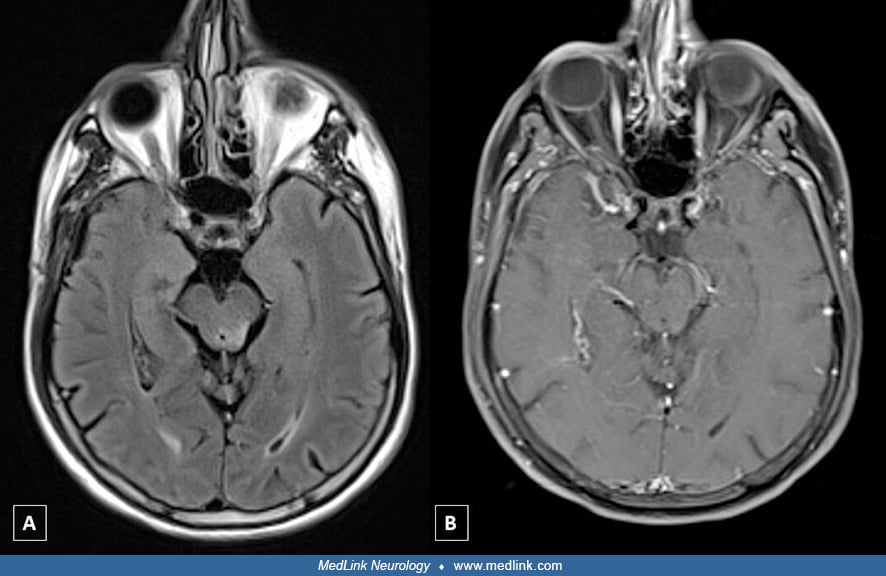
Enhancement of distal right internal auditory canal due to varicella-zoster virus (MRI)
Brain magnetic resonance image showing increased enhancement in the distal right internal auditory canal (arrow). (Source: Chou CC, Lo YT, Su HC, Chang CM. Fear of falling as a potential complication of Ramsay Hunt syndrome in older adults: a case report. BMC Geriatr 2022;22[1]:901. Creative Commons Attribution 4.0 International [CC-BY 4.0] license, creativecommons.org/licenses/by/4.0.)
-

Media
Ramsay Hunt syndrome (1-year follow-up)
One year after the patient’s Ramsay Hunt syndrome episode. Her facial palsy was considerably improved, and she received a Grade II score on the House-Brackmann Facial Paralysis Scale. (Source: Chou CC, Lo YT, Su HC, Chang CM. Fear of falling as a potential complication of Ramsay Hunt syndrome in older adults: a case report. BMC Geriatr 2022;22[1]:901. Creative Commons Attribution 4.0 International [CC-BY 4.0] license, creativecommons.org/licenses/by/4.0. The image was cropped from the original.)
-

Media
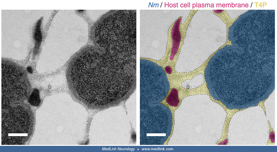
Working model of the mechanism of wetting-induced plasma membrane protrusions by N. meningitidis type IV pili fibers
N meningitidis produces type IV pili as a meshwork of fibers. On adhesion to the host endothelial cell, the high adhesiveness of type IV pili allows “one-dimensional” membrane wetting of the plasma membrane and, thus, remodeling of the membrane alongside type IV pili fibers. As the bacteria proliferate and aggregate extracellularly, plasma membrane protrusions remain attached to type IV pili fibers and end up embedded in a dense extracellular type IV pili meshwork. This complex structure gives microcolony enough mechanical coherence to resist blood flow–generated shear stress. (Source: Charles-Orszag A, Tsai FC, Bonazzi D, et al. Adhesion to nanofibers drives cell membrane remodeling through one-dimensional wetting. Nat Commun 2018;9[1]:4450. Creative Commons Attribution 4.0 International License, creativecommons.org/licenses/by/4.0.)


