Neuromuscular Disorders
Polymyositis, necrotizing autoimmune myositis, overlap-myositis, and myofasciitis
Feb. 28, 2024
MedLink®, LLC
3525 Del Mar Heights Rd, Ste 304
San Diego, CA 92130-2122
Toll Free (U.S. + Canada): 800-452-2400
US Number: +1-619-640-4660
Support: service@medlink.com
Editor: editor@medlink.com
ISSN: 2831-9125
Toll Free (U.S. + Canada): 800-452-2400
US Number: +1-619-640-4660
Support: service@medlink.com
Editor: editor@medlink.com
ISSN: 2831-9125
Nearly 3,000 illustrations, including video clips of neurologic disorders.
Every article is reviewed by our esteemed Editorial Board for accuracy and currency.
Full spectrum of neurology in 1,200 comprehensive articles.
Listen to MedLink on the go with Audio versions of each article.
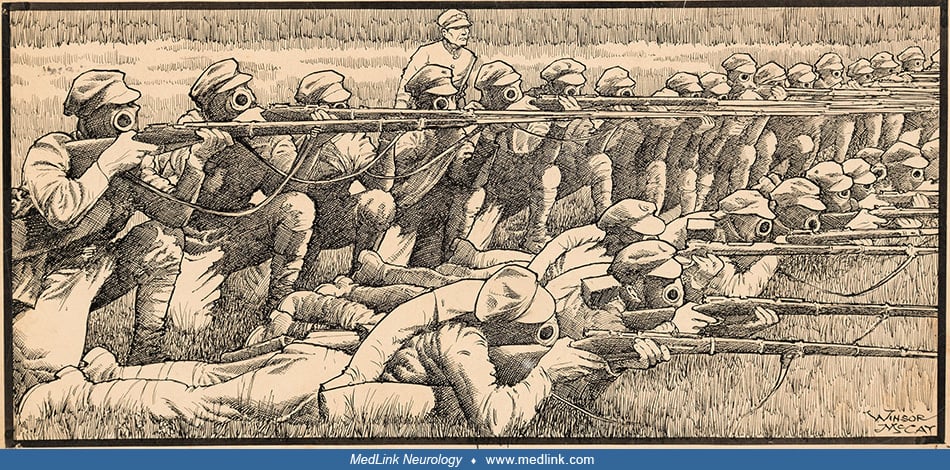
Color halftone printed in two colors after Scottish artist A. Kirkpatrick Maxwell (1884-1975), ca. 1917. The victim died at the end of the second day (40 hours after exposure). The bronchiole is filled with fibrin and pus cells, and its lining epithelium has been destroyed. The inflammation has caused a characteristic ring of hemorrhage in the tissues around the bronchial tube, and infection is beginning to appear in the alveoli nearest to these inflamed tissues. There is no generalized pulmonary edema. The pathological changes in the bronchioles and in the alveoli are, therefore, in sharp contrast with those caused by phosgene. As infection spreads into the lung tissues, patches of septic bronchopneumonia and small abscesses develop, and these often excite a surrounding inflammatory edema. If the patient lives, his bronchial mucous membrane is slowly regenerated; and during this time, he is subject to reflex spasms of coughing or even to a protracted bronchitis. (Source: Wellcome Collection, London, UK. Wellcome Library no. 571919i. Creative Commons Attribution 4.0 International [CC BY 4.0] license, creativecommons.org/licenses/by/4.0.)