Infectious Disorders
Central nervous system infection with Rickettsia species and related organisms
Jul. 03, 2024
MedLink®, LLC
3525 Del Mar Heights Rd, Ste 304
San Diego, CA 92130-2122
Toll Free (U.S. + Canada): 800-452-2400
US Number: +1-619-640-4660
Support: service@medlink.com
Editor: editor@medlink.com
ISSN: 2831-9125
Toll Free (U.S. + Canada): 800-452-2400
US Number: +1-619-640-4660
Support: service@medlink.com
Editor: editor@medlink.com
ISSN: 2831-9125
Nearly 3,000 illustrations, including video clips of neurologic disorders.
Every article is reviewed by our esteemed Editorial Board for accuracy and currency.
Full spectrum of neurology in 1,200 comprehensive articles.
Listen to MedLink on the go with Audio versions of each article.
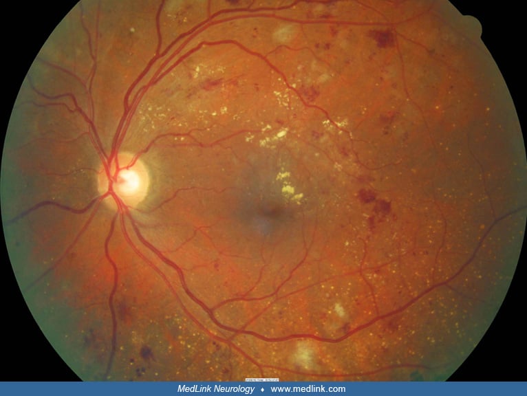
Dilated and tortuous retinal veins are seen along with scattered flame-shaped hemorrhages. The white patch temporal to the optic disc suggests secondary cilioretinal artery occlusion that occurred after the central retinal vein occlusion. (Contributed by Dr. Parin Hantrakul.)