Neuropharmacology & Neurotherapeutics
Rotigotine
Jul. 29, 2021
MedLink®, LLC
3525 Del Mar Heights Rd, Ste 304
San Diego, CA 92130-2122
Toll Free (U.S. + Canada): 800-452-2400
US Number: +1-619-640-4660
Support: service@medlink.com
Editor: editor@medlink.com
ISSN: 2831-9125
Toll Free (U.S. + Canada): 800-452-2400
US Number: +1-619-640-4660
Support: service@medlink.com
Editor: editor@medlink.com
ISSN: 2831-9125
Nearly 3,000 illustrations, including video clips of neurologic disorders.
Every article is reviewed by our esteemed Editorial Board for accuracy and currency.
Full spectrum of neurology in 1,200 comprehensive articles.
Listen to MedLink on the go with Audio versions of each article.
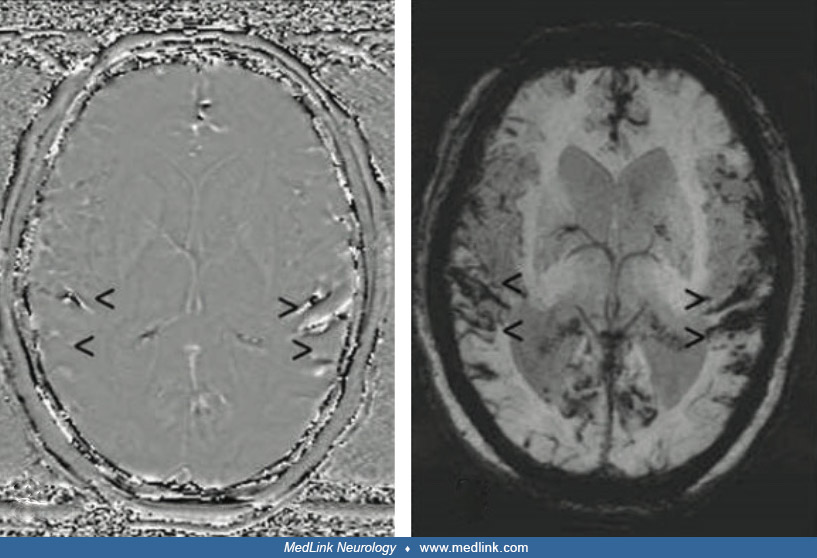
Acute cortical subarachnoid hemorrhage on (b) axial FLAIR, (c) axial susceptibility-weighted, and (d) axial diffusion-weighted images (arrow). (a, e) Enlarged perivascular spaces on axial T2-weighted images (arrow). (b, f) Focal gliosis on axial FLAIR (arrowhead). (c, g) Multiple microbleeds, cortical superficial siderosis, and residual atypical intracerebral bleeding in the left frontal area (arrowhead) on axial susceptibility-weighted images. (Source: Weidauer S, Neuhaus E, Hattingen E. Cerebral superficial siderosis: etiology, neuroradiological features and clinical findings. Clin Neuroradiol 2023;33[2]:293-306. Creative Commons Attribution 4.0 International [CC BY 4.0] license, creativecommons.org/licenses/by/4.0.)