Neuro-Ophthalmology & Neuro-Otology
Third nerve palsy
Nov. 22, 2024
MedLink®, LLC
3525 Del Mar Heights Rd, Ste 304
San Diego, CA 92130-2122
Toll Free (U.S. + Canada): 800-452-2400
US Number: +1-619-640-4660
Support: service@medlink.com
Editor: editor@medlink.com
ISSN: 2831-9125
Toll Free (U.S. + Canada): 800-452-2400
US Number: +1-619-640-4660
Support: service@medlink.com
Editor: editor@medlink.com
ISSN: 2831-9125
Nearly 3,000 illustrations, including video clips of neurologic disorders.
Every article is reviewed by our esteemed Editorial Board for accuracy and currency.
Full spectrum of neurology in 1,200 comprehensive articles.
Listen to MedLink on the go with Audio versions of each article.
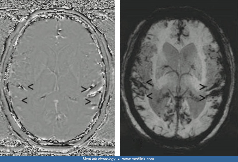
MRI sequences from a 61-year-old man with superficial siderosis due to ongoing hemorrhage from a melanoma metastasis in the right frontal cortex. In T2*-GRE (f) and susceptibility-weighted imaging (b, g), superficial siderosis is revealed by dark rims on the surface of affected structures, eg, the mesencephalon (arrow), with susceptibility-weighted imaging being more sensitive. Minimum intensity projections of susceptibility-weighted images (c, h) further enhance the conspicuousness of superficial siderosis. In addition, a filtered phase image of susceptibility-weighted imaging (a) can be used to distinguish paramagnetic (hemorrhage/iron, dark here) from diamagnetic substances (calcification, bright here), as they have opposite signal intensities. In general, susceptibility effects are more pronounced on images acquired at 3T (f-j) than on images acquired at 1.5T (a-e). Whereas at 3T a hypointense rim around the mesencephalon is seen in T2-weighted and FLAIR images (i, j; arrow), superficial siderosis is almost undetectable at 1.5T in T2-weighted and FLAIR images (d, e; arrow). (a-e) 1.5T; (a-c) susceptibility-weighted imaging, TR/alpha 52ms/20°, 4 echoes TE1 = 12 ms, deltaTE = 11 ms; (d) T2, TR/TE/alpha = 5762 ms/110 ms/90°; (e) FLAIR, TR/TE/TI/alpha = 11000 ms/140 ms/2800 ms/90°; (f-j) 3 T; (f) T2*-GRE, TR/TE/alpha=631ms/20ms/20°; (g, h) susceptibility-weighted imaging, TR/TE/alpha=27ms/20ms/15°; (i) T2, TR/TE/alpha=4980ms/92ms/150°, (j) FLAIR: TR/TE/TI/alpha = 8500 ms/81 ms/2440 ms/150°. TR repetition time, TE echo time, TI inversion time, α flip angle.
Abbreviation: T2*-GRE multi-echo gradient recalled echo T2*-weighted imaging (Tang MY, Chen TW, Zhang XM, Huang XH. GRE T2∗-weighted MRI: principles and clinical applications. Biomed Res Int 2014;2014:312142).
(Source: Weidauer S, Neuhaus E, Hattingen E. Cerebral superficial siderosis: etiology, neuroradiological features and clinical findings. Clin Neuroradiol 2023;33[2]:293-306. Creative Commons Attribution 4.0 International [CC BY 4.0] license, creativecommons.org/licenses/by/4.0.)