Developmental Malformations
Dandy-Walker syndrome
Jan. 19, 2024
MedLink®, LLC
3525 Del Mar Heights Rd, Ste 304
San Diego, CA 92130-2122
Toll Free (U.S. + Canada): 800-452-2400
US Number: +1-619-640-4660
Support: service@medlink.com
Editor: editor@medlink.com
ISSN: 2831-9125
Toll Free (U.S. + Canada): 800-452-2400
US Number: +1-619-640-4660
Support: service@medlink.com
Editor: editor@medlink.com
ISSN: 2831-9125
Nearly 3,000 illustrations, including video clips of neurologic disorders.
Every article is reviewed by our esteemed Editorial Board for accuracy and currency.
Full spectrum of neurology in 1,200 comprehensive articles.
Listen to MedLink on the go with Audio versions of each article.
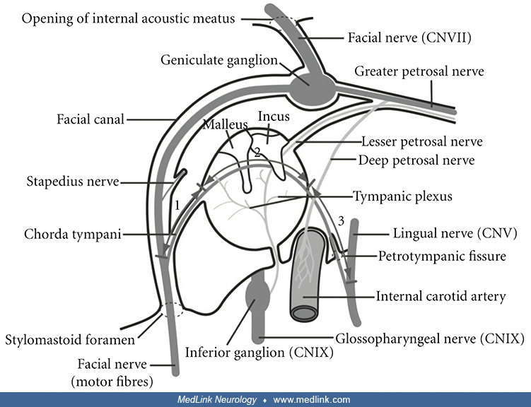
A view of the right-sided middle ear space with the external ear canal, eardrum (tympanic membrane) and hearing bones (ossicles) removed, showing the course of the chorda tympani nerve. The observer is looking at the medial wall of the middle ear, as seen from a lateral viewpoint. The facial nerve is seen to pass horizontally in a path superior to the middle ear and then turn in an inferior direction and pass vertically posterior to the middle ear. The Eustachian tube extends anteriorly and is directed inferiorly as its path towards the throat (nasopharynx) extends further from the middle ear. The jugular fossa is inferior to the middle ear space. (Source: Illustration by Henry Vandyke Carter [1831-1897]. Gray H, Lewis WH. Anatomy of the Human Body. Twentieth edition. Philadelphia and New York: Lea & Febiger, 1918. Public domain.)