Neuro-Oncology
Radiation myelopathy
Oct. 17, 2023
MedLink®, LLC
3525 Del Mar Heights Rd, Ste 304
San Diego, CA 92130-2122
Toll Free (U.S. + Canada): 800-452-2400
US Number: +1-619-640-4660
Support: service@medlink.com
Editor: editor@medlink.com
ISSN: 2831-9125
Toll Free (U.S. + Canada): 800-452-2400
US Number: +1-619-640-4660
Support: service@medlink.com
Editor: editor@medlink.com
ISSN: 2831-9125
Nearly 3,000 illustrations, including video clips of neurologic disorders.
Every article is reviewed by our esteemed Editorial Board for accuracy and currency.
Full spectrum of neurology in 1,200 comprehensive articles.
Listen to MedLink on the go with Audio versions of each article.
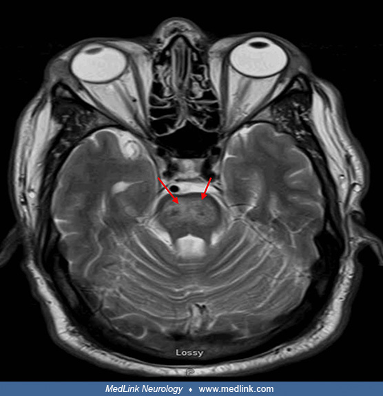
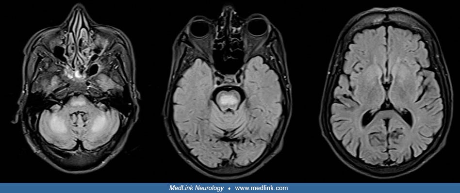
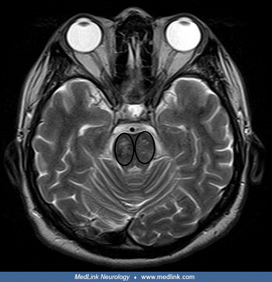
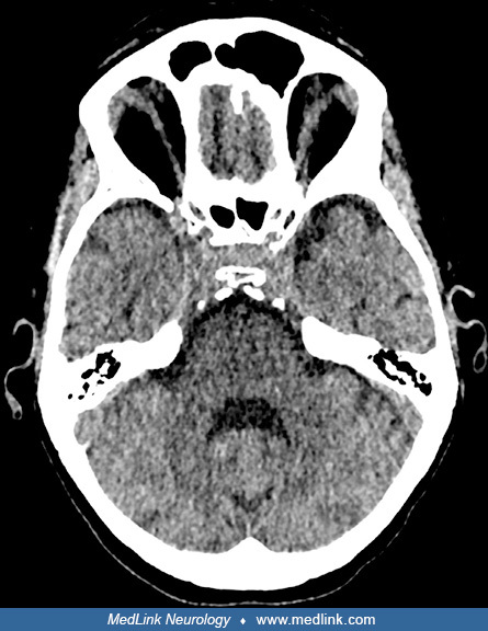
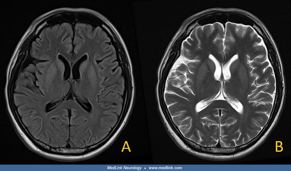
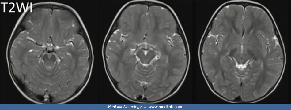
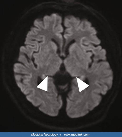
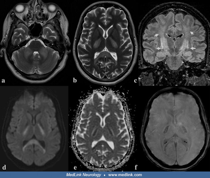
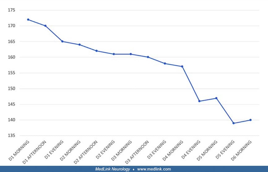
CT scan of the head showing scattered areas of reduced density in the subcortical white matter, including the frontal lobes and right parietal lobe, consistent with extrapontine myelinolysis. This 40-year-old woman with chronic alcoholism presented with intractable headache for 3 days and progressively worsening unsteady gait requiring a wheelchair to ambulate. Electrolyte levels were normal. Central pontine myelinolysis and extrapontine myelinolysis were attributed to alcohol withdrawal. (Source: Jamil M, Salam A, Joseph Benher BM, Rehman S, Jamil J, Suleyman G. A case of alcohol withdrawal-induced central and extrapontine myelinolysis. Cureus 2023;15[7]:e41640. Creative Commons Attribution 4.0 International [CC BY 4.0] license, creativecommons.org/licenses/by/4.0.)